
How to differentiate clinically between LTB (croup) and epiglottitis
Which of the following best differentiates, by history, LTB (croup) from epiglottitis?
The answer is C.
Croup, laryngeotrachiobronchitis, a viral illness with subglottic involvement, typically exhibits the symptoms of inspiratory stridor only after 12 to 24 hours of coryza, typical of a viral “cold.” Epiglottitis, which is a much more serious condition, begins suddenly. Epiglottitis is caused by bacterial infection with supraglottic involvement, classically by H.influenzae but also by S.aureus and Corynebacterium diphtheriae.
 Although epiglottitis is characterized more by high fever than is croup, this appears to be a weak factor on which to base a preliminary diagnosis. Whereas croup virtually always includes a cough, epiglottitis rarely
does so. Epiglottitis typically includes dysphagia while croup does not. The child with croup is comfortable in all positions, whereas the patient with epiglottitis will be sitting forward with the mouth open. Both conditions are characterized by inspiratory dyspnea. Croup is benign and epiglottitis is potentially critical.
- (A) Temperature
- (B) Presence of inspiratory stridor
- (C) Length of time from onset to defining symptom
- (D) Lung field auscultation
- (E) Presence or absence of dyspnea
The answer is C.
Croup, laryngeotrachiobronchitis, a viral illness with subglottic involvement, typically exhibits the symptoms of inspiratory stridor only after 12 to 24 hours of coryza, typical of a viral “cold.” Epiglottitis, which is a much more serious condition, begins suddenly. Epiglottitis is caused by bacterial infection with supraglottic involvement, classically by H.influenzae but also by S.aureus and Corynebacterium diphtheriae.

does so. Epiglottitis typically includes dysphagia while croup does not. The child with croup is comfortable in all positions, whereas the patient with epiglottitis will be sitting forward with the mouth open. Both conditions are characterized by inspiratory dyspnea. Croup is benign and epiglottitis is potentially critical.
Sesamoid bones, Functions and mechanism of action.
The sesamoid bone is a small rounded bone embedded within a tendon that usually passes over a joint (Sesamoid bones are the bones not connected to any other bone).The largest sesamoid bobe is the patella.

Sesamoid bones functions probably are to modify pressure, to diminish friction, and occasionally to alter the direction of a muscle pull.Sesamoid bones also prevent the tendon from flattening into the joint as tension increases and therefore also maintain a more consistent moment arm through a variety of possible tendon loads.
Sesamoid bones can be found in the knee, hand and foot...

Latin........."ossa sesamoidea"

Sesamoid bones functions probably are to modify pressure, to diminish friction, and occasionally to alter the direction of a muscle pull.Sesamoid bones also prevent the tendon from flattening into the joint as tension increases and therefore also maintain a more consistent moment arm through a variety of possible tendon loads.
Sesamoid bones can be found in the knee, hand and foot...

Sesamoid bones at the distal end of the first metatarsal bone of the foot.
Sesamoids act like pulleys. They provide a smooth surface over which the tendons slide, so they increase the ability of the tendons to transmit muscle forces. The sesamoids in the forefoot also assist with weightbearing and help elevate the bones of the big toe.
Associated conditions result in intestinal intussusception in children

- A) colon polyp
- B) Meckel's diverticulum
- C) lymphoma
- D) parasite infection
- E) foreign body
The answer is C.
Intussusception is the most common cause of intestinal obstruction in the first 2 years of life. It is more common in males than in females. In most cases (85%) the cause is not apparent. Associated conditions that can result in intussusception include :-polyps, Meckel's diverticulum, Henoch–Schonlein purpura, lymphoma, lipoma, parasites, foreign bodies, and viral enteritis with hypertrophy of Peyer patches.
Intussusception of the small intestine occurs in patients with celiac disease and cystic fibrosis—related to the bulk of stool in the terminal ileum. Henoch–Schonlein purpura may also cause isolated small-bowel intussusception. In children older than 6 years, lymphoma is the most common cause. Intermittent small-bowel intussusception is a rare cause of recurrent abdominal pain.
Hay WW Jr, Levin MJ, Sondheimer JM, et al., eds. Current pediatric diagnosis & treatment, 18th ed. New York: McGraw-Hill; 2007:616–617
Burns Injury and Burns Degrees
 | |
| example of 2nd dgree burn |
A burn is a part of traumatology that is caused by heat, radiation, chemicals or electricity. Burns affect usually the skin (epidermis and dermis).Affection of deeper tissues, such as muscle, bone, and blood vessels may be fatal
 |
1-Outer skin layer 2-Middle skin layer 3-Deep skin layer 4-First degree burn 5-Second degree burn 6-Third degree burn |
 |
| 1st degrre burn |
1st degree burn usullay has no complication and healed in about 1 week.
2- 2nd Degree Burns ; this type of burn affects tha dermis layer and may be subdivided into superficial partial thickness and deep partial thickness.
These second-degree burns are characterized by extreme pain and appears as wet, red, swollen and blistered areas. healing time 2-3 weeks and complications like Local infection/cellulitis, Scarring, Contractures may be seen.

*dry and leathery skin
*black, white, brown, or yellow skin
*swelling
*lack of pain because nerve endings have been destroyed
4- 4th Degree Burns ;are life threatening injury where the burn Extends through skin, subcutaneous tissue and into underlying muscle and bone, the affect limb usually amputated.
Treating pressure ulcers

- A) application of povidone-iodine gauze two times per day
- B) application of hydrogen peroxide 3 times per day
- C) systemic antibiotics for 7 to 10 days
- D) keeping the area clean and dry until granulation tissue forms
- E) surgical debridement
The answer is E. (Surgical débridement)
When treating this pressure ulcer, it is important to maintain a moist environment while keeping the surrounding skin dry. This can be accomplished by loosely packing the ulcer with saline-moistened gauze. Topical antimicrobials such as silver sulfadiazine cream may be helpful in ulcers that appear infected. Topical antiseptics such as povidone-iodine or hydrogen peroxide should be not be used in the treatment of pressure ulcers. Systemic antibiotics should be reserved for serious infections (e.g., bacteremia, osteomyelitis). A 2-week trial of topical antimicrobials may be considered for ulcers that do not appear infected but are not improving. Although most patients are successfully managed without surgery, procedures may be appropriate in patients whose quality of life would be markedly improved by rapid wound closure. Stage 3 and 4 ulcers with necrotic tissue should be débrided. Ulcers with minimal exudate that are not infected can be covered with an occlusive dressing to promote autolytic débridement. Ulcers with thick exudate, slough, or loose necrotic tissue should undergo mechanical débridement. Options include wet-to-dry dressings, hydrotherapy, wound irrigation, and scrubbing the wound with gauze. Ulcers with evidence of cellulitis or deep infection should undergo sharp débridement with a scalpel or scissors. Ulcers with a thick eschar or extensive necrotic tissue should undergo sharp débridement as well. However, a thick, dry eschar covering a heel ulcer should generally be left intact. Patients without access to surgical inter-ventions (such as in a long-term care setting) or those who may not be acceptable surgery candidates can be treated with enzymatic débriding agents. Wound débridement should stop once necrotic tissue has been removed and granulation tissue is present.
Illustration of Lumbar Hernia

These occur more commonly in males and are twice as common on the left than the right side. Patients are usually between 50 to 70 years old. These hernias can occur anywhere within the lumbar region but are more common through the superior lumbar triangle (of Grynfeltt-Lesshaft)
So, Lumbar hernia has 2 anatomical types:
1-Petit's hernia: a hernia through Petit's triangle (inferior lumbar triangle). It is named after French surgeon Jean Louis Petit (1674–1750).
2-Grynfeltt's hernia: a hernia through Grynfeltt-Lesshaft triangle (superior lumbar triangle). It is named after physician Joseph Grynfeltt (1840–1913).

**Patients with lumbar hernia are usually asymptomatic but may complain of backache, flank pain or a dragging sensation. These hernias have a natural history of a gradual increase in size over time.

At this stage include the differential considerations includes lipoma, soft tissue tumors, hematoma or abscess.
Illustrated Cephalohematoma Vs Caput succedaneum
 Cephalohematoma is a collection of blood under the periosteum of a skull bone "very tough tissue covering that encapsulates bones"
Because of its location, it is impossible for cephalohematoma to cross suture lines. If more than one bone is affected, there will be a separation between the two areas at the suture line as seen in this photo at the left where the sagittal suture separates the bilateral parietal cephalohematomas.
Unlike cephalohematoma; A caput succedaneum is caused by the mechanical trauma of the initial portion of scalp pushing through a narrowed cervix. The swelling may be on any portion of the scalp, may cross the midline (as opposed to a cephalhematoma), and may be discolored because of slight bleeding in the area. There may also be molding of the head, which is common in association with a caput succedaneum.
 A cephalohematoma in a 1-week-old newborn with a right parietal bump by vacuum extractor . A plain skull X-ray lateral view revealing the linear skull fracture on the right parietal area .
Appearance of Sunset sign in infant eyes

The sclera are visible between the upper eyelid and the iris,Sunsetting sign is seen usually in hydrocephalus due to loss of upward conjugate gaze caused by raised intracranial pressure (ICP)

However, it can also be transiently elicited in healthy infants up to 7 months of age by changes of position or removal of light (benign setting-sun phenomenon). The benign form might represent immaturity of the reflex systems controlling eye movements.
When persistent, this sign is one of the most frequent markers of elevated intracranial pressure, appearing in 40% of children with hydrocephalus (whatever the cause of Hydrocephalus as obstructive, communicating, Dandy-Walker anomaly/syndrome) and in 13% of patients with ventriculoperitoneal shunt dysfunction. It is an earlier sign of hydrocephalus than enlarged head circumference, full fontanelle, separation of sutures, irritability or vomiting. Consequently, this sign is a valuable early warning of an entity requiring prompt neuroimaging and urgent surgical intervention.
Synonyms
baby, Downward deviation of eyes, Downward Ocular Deviation, EYE DEVIATION DOWNWARD, infant, Infant (person), Infant child, Infant child (person), Infants, Manifestation Neurologic, Manifestation Neurological, MANIFESTATIONS NEUROL, Manifestations Neurologic, Manifestations Neurological, neural manifestation, NEUROL MANIFESTATION, NEUROL MANIFESTATIONS, NEUROL SIGNS SYMPTOMS, neurologic manifestation, Neurologic Manifestations, Neurologic manifestations of general diseases, Neurologic Signs and Symptoms, Neurological Manifestation, Neurological Manifestations, Sunset Sign
Lines on anterior Abdominal Wall
This photo shows surface anatomy of some lines on the anterior abdominal wall like:
 1-McBurney's point is halfway between the umbilicus and the ASIS ( anterior superior illiac spine ) and it is a common location where surgeons use for an incision to remove the appendix.
2-The linea alba is a fibrous structure that runs down the midline of the abdomen and seperates the left and right rectus abdominus muscles.
3-The arcuate line demarcates the lower limit of the posterior layer of the rectus sheath.
4-The inguinal ligament is a band running from the pubic tubercle to the anterior superior iliac spine, its anatomy is very important for operating on hernia patients.
This ligament passes between two bony points of the hip bone, the anterior superior iliac spine laterally and the pubic tubercle medially. It has an expanded medial end, the lacunar ligament.
The inguinal ligament is the thickened, recurved free inferior border of the external oblique muscle. It forms the floor of the inguinal canal along which passes the spermatic cord in the male or the round ligament of the uterus in the female.

5-The linea semilunaris is a curved tendinous line placed one on either side of the rectus abdominus and corresponds with the lateral border of the rectus muscle.

2-The linea alba is a fibrous structure that runs down the midline of the abdomen and seperates the left and right rectus abdominus muscles.
3-The arcuate line demarcates the lower limit of the posterior layer of the rectus sheath.
4-The inguinal ligament is a band running from the pubic tubercle to the anterior superior iliac spine, its anatomy is very important for operating on hernia patients.
This ligament passes between two bony points of the hip bone, the anterior superior iliac spine laterally and the pubic tubercle medially. It has an expanded medial end, the lacunar ligament.
The inguinal ligament is the thickened, recurved free inferior border of the external oblique muscle. It forms the floor of the inguinal canal along which passes the spermatic cord in the male or the round ligament of the uterus in the female.

5-The linea semilunaris is a curved tendinous line placed one on either side of the rectus abdominus and corresponds with the lateral border of the rectus muscle.
Scaphoid fractures overview

Anatomic snuffbox tenderness is a highly sensitive test for scaphoid fracture, whereas scaphoid compression pain and tenderness of the scaphoid tubercle tend to be more specific. Initial radiographs in patients suspected of having a scaphoid fracture should include anteroposterior, lateral, oblique, and scaphoid wrist views.

RADIOGRAPHY :
 |
| scaphoid view |
Initial radiographs do not always detect scaphoid fractures. In one prospective trial,8 the sensitivity of initial radiographs was 86 percent. However, a great deal of variability in the sensitivities (higher and lower) of radiographs is found in the literature. Nondisplaced fractures of this bone are known to be difficult to see on initial radiographs. In these cases, one treatment option includes placing the patient in a cast and performing a follow-up physical examination and repeat radiography in two weeks. Recent improvements in technology may allow alternate approaches in this situation.

(Left) This x-ray shows a scaphoid fracture fixed in place with a screw. (Right) This x-ray was taken 4 months after surgery. The fracture of the scaphoid is healed.
X-ray Osgood-Schlatter disease
Osgood Schlatters disease is a very common cause of knee pain in both children and young athletes usually between the ages of 10 and 16. It occurs due to a period of rapid growth, combined with a high level of sporting activity.
* Normal x-ray findings do not exclude the disease, which is diagnosed clinically
* Radiographs have Limited role "Clinical diagnosis" and are usually obtained to exclude other causes of pain
* Conventional radiography
o Not helpful if tubercle has not calcified (usually around 9 [girls]-11 [boys] years of age)
o Best seen on lateral knee
o Irregular ossification or fragmentation of tibial tubercle..... Separated from remainder of tibial tubercle
o Soft tissue swelling
o Calcification in or thickening of the patellar tendon

Imaging Findings
* Normal x-ray findings do not exclude the disease, which is diagnosed clinically
* Radiographs have Limited role "Clinical diagnosis" and are usually obtained to exclude other causes of pain
* Conventional radiography
o Not helpful if tubercle has not calcified (usually around 9 [girls]-11 [boys] years of age)
o Best seen on lateral knee
o Irregular ossification or fragmentation of tibial tubercle..... Separated from remainder of tibial tubercle
o Soft tissue swelling
o Calcification in or thickening of the patellar tendon

Lateral radiograph of the knee demonstrating fragmentation of the tibial tubercle with overlying soft tissue swelling.
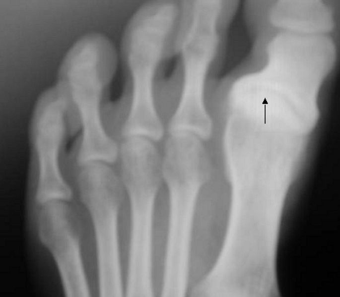
Hallux varus in X-ray

The condition of Hallux varus deformity has various degrees of severity and causes. Hallux varus most commonly caused by rupture of the lateral collateral ligament at the MTP joint following a surgical procedure "as a previous bunion surgery" or trauma but it can also be due to removal of sesamoid bones, arthritis, or congenital deformity.
A case of Alopecia areata
A 37y old man presents with rapid hair loss that has occurred over the last few weeks. He reports that his father had a similar condition. The most likely diagnosis is :

The answer is A. "Alopecia areata"
Alopecia areata is associated with sudden hair loss that occurs in round patches. The patches are well circumscribed, not associated with scarring or inflammation and the patients have no other symptoms. The most common area affected is the scalp; but, the condition may also affect the eyebrows or beard. Alopecia areata usually affects children and young adults and is recurrent.
 A pathognomonic sign for alopecia areata is the “exclamation point” hair, which is wide distally and narrower at the base. These hairs are often found at the periphery of a patch of hair loss. Hair that regrows in the area of alopecia areata is in many cases white. Nail pitting may be also present. The treatment consists of injection of intralesional steroids and topical steroids. Most experience complete regrowth of hair.
- A) alopecia areata
- B) tinea capitis
- C) androgenic alopecia
- D) secondary syphilis
- E) trichotillomania
The answer is A. "Alopecia areata"
Alopecia areata is associated with sudden hair loss that occurs in round patches. The patches are well circumscribed, not associated with scarring or inflammation and the patients have no other symptoms. The most common area affected is the scalp; but, the condition may also affect the eyebrows or beard. Alopecia areata usually affects children and young adults and is recurrent.

Koplik’s spots as described
 This was a patient who presented with Koplik’s spots on palate due to pre-eruptive measles on day 3 of the illness. Measles is an acute, highly communicable viral disease with fever, conjunctivitis, coryza, cough, and Koplik spots. Koplik spots are small, red, irregularly-shaped spots with blue-white centers found on the mucosal surface of the oral cavity.
The spots as described by Koplik in 1896:
If we look in the mouth at this period we see a redness of the fauces; perhaps, not in all cases, a few spots on the soft palate. On the buccal mucous membrane and the inside of the lips, we invariably see a distinct eruption. It consists of small, irregular spots, of a bright red colour. In the centre of each spot, there is noted, in strong daylight, a minute bluish white speck. These red spots, with accompanying specks of a bluish colour, are absolutely pathognomonic of beginning measles, and when seen can be relied upon as the forerunner of the skin eruption.
About head lice infestations
Which of the following statements is true regarding head lice infestations?
The answer is: ( A ).
 Head lice are thought to be the most common type and are developing resistance to commonly used pediculicides. Every year, between 6 million and 12 million people in the United States, primarily children 3 to 10 years of age, are infested with head lice. Girls are at greater risk because they have more frequent head-to-head contact.
Head lice affect people of all socioeconomic status. Head lice are obligate parasites that live on human skin and survive on human blood. No other animal is affected. Head lice die if they are away from the host head for more than 2 days.
Lice are wingless and cannot jump, but they climb quickly from hair to hair when the hair is dry. Lice move slowly on wet hair and can be removed more easily with a gloved hand or a fine-toothed comb. The adult female louse lays 7 to 10 eggs daily that attach to human hair with a gluelike, water-soluble substance. By 7 to 10 days, a nymph emerges from the egg and is close enough to the scalp to obtain its first meal of blood. Adult lice, after the 7- to 10-day period of molting stages, live about 30 days. Infested people usually have no more than 10 to 12 live head lice at a time, but can harbor hundreds of eggs and nits. Those affected describe itching and a sensation of “something crawling on my head"? Scratching may cause excoriations and secondary infection. Most infestations are asymptomatic.
All household members with active infestation should be treated simultaneously. For children younger than 2 years, there is no recommended pediculicide; therefore, treatment consists of manual removal only. Lice that remain active 8 to 12 hours after treatment may require an alternative agent. Itching may persist for up to 10 days after successful treatment and should not be mistaken for treatment failure. The Centers for Disease Control and Prevention recommend that all clothing and bedding in contact with the infested person during the 2 days before application of the pediculicide be laundered in hot water and machine dried using a hot dry cycle. All nonwashable items should be quarantined in a plastic bag for 2 weeks. Combs and brushes should be disinfected with hot water or alcohol.
- A) Females are more likely affected.
- B) Retreatment with pyrethrin is rarely needed.
- C) Head lice can live off the body up to 1 week.
- D) Low socioeconomic children are more likely affected.
- E) Dogs are a common vector for head lice.
The answer is: ( A ).

Head lice affect people of all socioeconomic status. Head lice are obligate parasites that live on human skin and survive on human blood. No other animal is affected. Head lice die if they are away from the host head for more than 2 days.
Lice are wingless and cannot jump, but they climb quickly from hair to hair when the hair is dry. Lice move slowly on wet hair and can be removed more easily with a gloved hand or a fine-toothed comb. The adult female louse lays 7 to 10 eggs daily that attach to human hair with a gluelike, water-soluble substance. By 7 to 10 days, a nymph emerges from the egg and is close enough to the scalp to obtain its first meal of blood. Adult lice, after the 7- to 10-day period of molting stages, live about 30 days. Infested people usually have no more than 10 to 12 live head lice at a time, but can harbor hundreds of eggs and nits. Those affected describe itching and a sensation of “something crawling on my head"? Scratching may cause excoriations and secondary infection. Most infestations are asymptomatic.
Prior to treatment, live lice can be identified under a magnifying glass, which is best done when the hair is wet. After infestation is confirmed, treatment consists of application of a pediculicidal agent to the hair, followed by mechanical removal.
The hair should not be washed for 2 to 3 days after the pediculicide is applied. Thorough physical removal of lice and nits with a sturdy, fine-tooth comb is recommended for several days after application because no pediculicide guarantees total eradication of lice. First-line topical agents containing pyrethrins or permethrin are available over the counter and are relatively nontoxic. Retreatment is advised 7 to 10 days after the first application of pyrethrins. Because permethrin remains active for a longer period, retreatment generally is not necessary. Permethrin 5% cream is available by prescription for use in resistant cases. Alternative agents include lindane or malathion. Lindane has been shown to have limited success and is systemically absorbed, so its use is now considered second-line. Malathion was recently labeled by the U.S. Food and Drug Administration (FDA) for the treatment of head lice and is available in a lotion that is left on the head for 8 to 12 hours. Oral agents include ivermectin and trimethoprim–sulfamethoxizole. Resistance is possible with any treatment because of reduced susceptibility or incorrect use of the medication. All household members with active infestation should be treated simultaneously. For children younger than 2 years, there is no recommended pediculicide; therefore, treatment consists of manual removal only. Lice that remain active 8 to 12 hours after treatment may require an alternative agent. Itching may persist for up to 10 days after successful treatment and should not be mistaken for treatment failure. The Centers for Disease Control and Prevention recommend that all clothing and bedding in contact with the infested person during the 2 days before application of the pediculicide be laundered in hot water and machine dried using a hot dry cycle. All nonwashable items should be quarantined in a plastic bag for 2 weeks. Combs and brushes should be disinfected with hot water or alcohol.
Tongue`s surface in Prolonged antibiotic use

- A) Prolonged antibiotic use
- B) Sjögren's syndrome
- C) Addison's disease
- D) Chronic gastroesophageal reflux
- E) Malignant melanoma
The answer is A.
(Black tongue) actually ...Black hairy tongue results from hyperplasia of the filiform papillae with deposition of keratin on the surface. The condition causes the tongue to have a dark, velvety, hairlike appearance.Associated conditions include smoking, consumption of coffee, prolonged use of antibiotics, and possibly acquired immunodeficiency syndrome. Treatment involves using a toothbrush to scrape off the excess keratin that forms on the tongue's suface.
Photo illustration of Allen’s Test
Photo illustration of Allen’s Test

+ve Allen test :
When you occlude the radial artery for several minutes and compares the hand color to the other hand and you find that there is no change in color------>This hand is said to have sufficient collateral circulation through the ulnar artery . |
| Step 1 occlusion |
 |
| Step 2 releasing |
Modified Allen’s test :
Step 1 occlusion : With the same idea, Ask the patient to clench the hand into a tight fist and then apply pressure to both the radial and ulnar arteries, and ask the patient to relax their hand .The hand should appear somewhat white & blanched (if not....you haven`t completely occluded the arteries with your fingers). Step 2 releasing : When you Release the occlusive pressure on the ulnar artery it will result in the hand regaining its colour "flushing of the hand within 5 to 15 seconds"------> the ulnar collateral circulation is adequate, and it`s called positive modified Allen’s test
The test can be repeated, this time releasing the pressure on the radial artery, to assess radial collateral circulation.
Subscribe to:
Posts (Atom)