Showing posts with label ORTHOPEDICS. Show all posts
Showing posts with label ORTHOPEDICS. Show all posts
Sesamoid bones, Functions and mechanism of action.
The sesamoid bone is a small rounded bone embedded within a tendon that usually passes over a joint (Sesamoid bones are the bones not connected to any other bone).The largest sesamoid bobe is the patella.

Sesamoid bones functions probably are to modify pressure, to diminish friction, and occasionally to alter the direction of a muscle pull.Sesamoid bones also prevent the tendon from flattening into the joint as tension increases and therefore also maintain a more consistent moment arm through a variety of possible tendon loads.
Sesamoid bones can be found in the knee, hand and foot...

Latin........."ossa sesamoidea"

Sesamoid bones functions probably are to modify pressure, to diminish friction, and occasionally to alter the direction of a muscle pull.Sesamoid bones also prevent the tendon from flattening into the joint as tension increases and therefore also maintain a more consistent moment arm through a variety of possible tendon loads.
Sesamoid bones can be found in the knee, hand and foot...

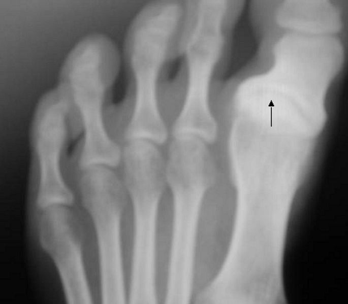
Sesamoid bones at the distal end of the first metatarsal bone of the foot.
Sesamoids act like pulleys. They provide a smooth surface over which the tendons slide, so they increase the ability of the tendons to transmit muscle forces. The sesamoids in the forefoot also assist with weightbearing and help elevate the bones of the big toe.
Scaphoid fractures overview

Anatomic snuffbox tenderness is a highly sensitive test for scaphoid fracture, whereas scaphoid compression pain and tenderness of the scaphoid tubercle tend to be more specific. Initial radiographs in patients suspected of having a scaphoid fracture should include anteroposterior, lateral, oblique, and scaphoid wrist views.

RADIOGRAPHY :
 |
| scaphoid view |
Initial radiographs do not always detect scaphoid fractures. In one prospective trial,8 the sensitivity of initial radiographs was 86 percent. However, a great deal of variability in the sensitivities (higher and lower) of radiographs is found in the literature. Nondisplaced fractures of this bone are known to be difficult to see on initial radiographs. In these cases, one treatment option includes placing the patient in a cast and performing a follow-up physical examination and repeat radiography in two weeks. Recent improvements in technology may allow alternate approaches in this situation.

(Left) This x-ray shows a scaphoid fracture fixed in place with a screw. (Right) This x-ray was taken 4 months after surgery. The fracture of the scaphoid is healed.
X-ray Osgood-Schlatter disease
Osgood Schlatters disease is a very common cause of knee pain in both children and young athletes usually between the ages of 10 and 16. It occurs due to a period of rapid growth, combined with a high level of sporting activity.
* Normal x-ray findings do not exclude the disease, which is diagnosed clinically
* Radiographs have Limited role "Clinical diagnosis" and are usually obtained to exclude other causes of pain
* Conventional radiography
o Not helpful if tubercle has not calcified (usually around 9 [girls]-11 [boys] years of age)
o Best seen on lateral knee
o Irregular ossification or fragmentation of tibial tubercle..... Separated from remainder of tibial tubercle
o Soft tissue swelling
o Calcification in or thickening of the patellar tendon

Imaging Findings
* Normal x-ray findings do not exclude the disease, which is diagnosed clinically
* Radiographs have Limited role "Clinical diagnosis" and are usually obtained to exclude other causes of pain
* Conventional radiography
o Not helpful if tubercle has not calcified (usually around 9 [girls]-11 [boys] years of age)
o Best seen on lateral knee
o Irregular ossification or fragmentation of tibial tubercle..... Separated from remainder of tibial tubercle
o Soft tissue swelling
o Calcification in or thickening of the patellar tendon
Lateral radiograph of the knee demonstrating fragmentation of the tibial tubercle with overlying soft tissue swelling.
Hallux varus in X-ray

The condition of Hallux varus deformity has various degrees of severity and causes. Hallux varus most commonly caused by rupture of the lateral collateral ligament at the MTP joint following a surgical procedure "as a previous bunion surgery" or trauma but it can also be due to removal of sesamoid bones, arthritis, or congenital deformity.
Subscribe to:
Posts (Atom)